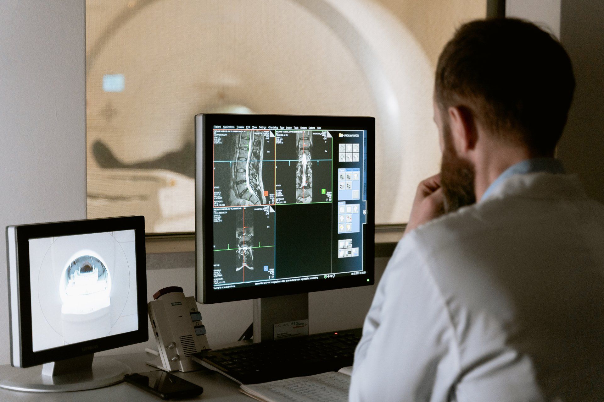
High Speed and High resolution imaging of the whole brain is essential to neurovascular research.
Doing these things individually is the standard, doing them together however is extremely difficult.
A new study by engineers at Duke University have solved this long-standing trade off. These engineers have developed a method to scan and image brain activity in real time with a high enough resolution which could reveal new insights into neurovascular diseases like stroke, dementia and even acute brain injuries.
This method, named “Ultrafast Photoacoustic Microscopy” or UFF-PAM relies on advancements in hardware and machine learning algorithms to upgrade current techniques. Allowing medical professionals to view imagery without sacrificing a full field of view of the patients’ brain. Improving speed and detail within this type of imaging has allowed scientists to observe how the brain responds to a stroke and how it begins to recover in the final stage.
Observing that blood vessels constrict immediately after a stroke and compress neighbouring vessels the team pinpointed the starting position of this constriction and tracked its movement through the brain. In the future scientists have planned to use the UFF-PAM to explore brain disease models such as dementia or Alzheimer’s disease.
They also believe that the tool can be used outside the brain to image organs such as the heart, liver and placenta.
"Mapping the brain efficiently has eluded us for years. With this step we are getting closer... imagine the remedies and answers we could find." Shivvy Jervis